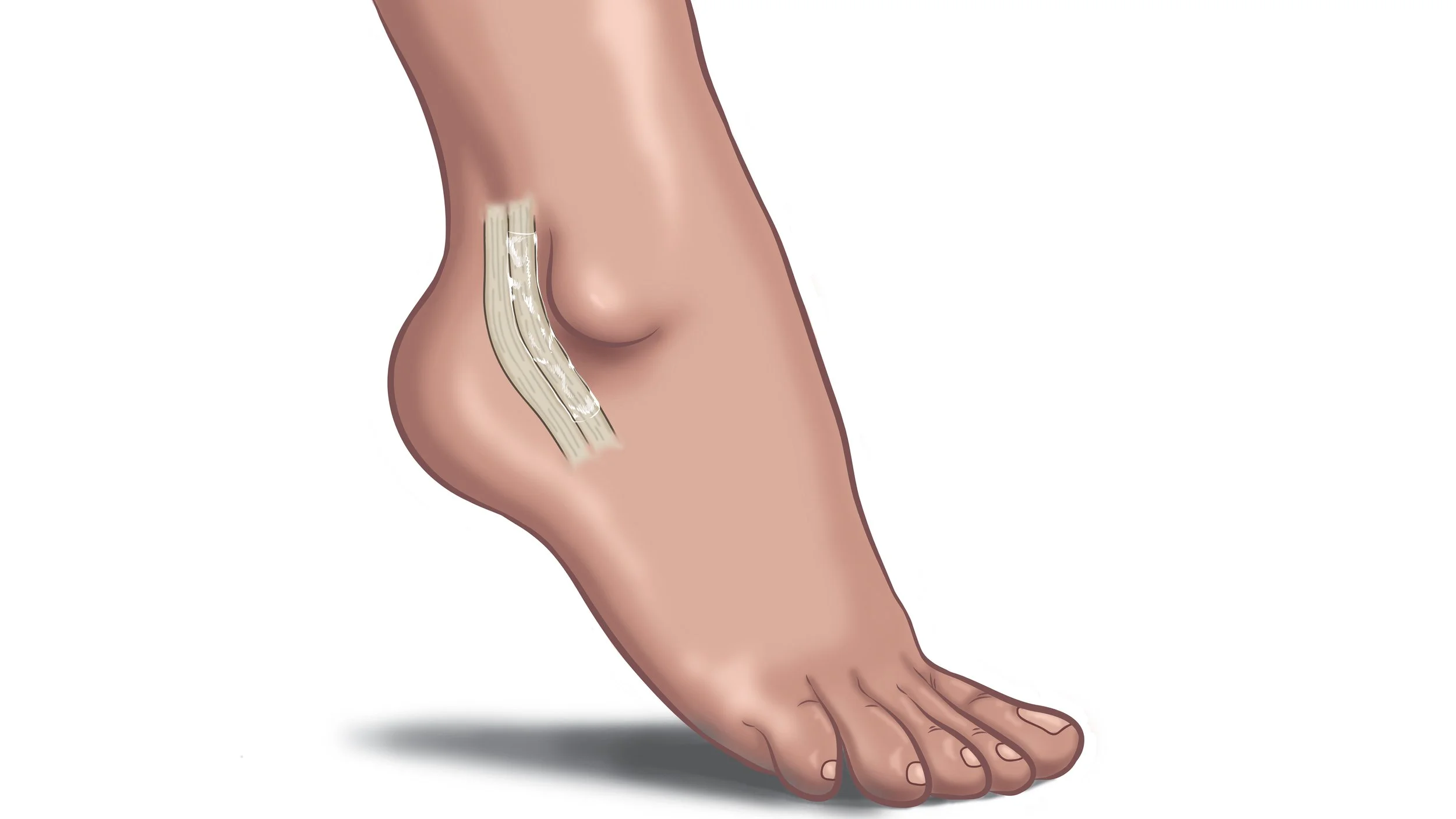

VersaWrap® is a medical device implant that provides a gliding surface for tendons and peripheral nerve in foot, ankle, and other lower extremity procedures
- Peroneal repairs
- Tarsal tunnel revisions
- Achilles’ procedures
- Anterior talo-fibular ligament (ATFL)
- Transfers

Examples of VersaWrap application in lower extremity procedures
Click the images to learn more
Peroneal tendon dislocation reconstruction
-
The patient is a 44-year-old active female who had a twisting injury while skiing 3 months prior to surgery. She experienced lateral ankle pain and snapping of the peroneal tendons during activity. The axial T2 MRI demonstrates a superior peroneal retinacular tear(SPR) with a split tear of the peroneus brevis.
-
Peroneal tendoscopy was performed followed by an open repair which revealed a denuded distal fibula.
VersaWrap was placed around the peroneal tendons prior to the final repair of the SPR.
-
The final repair allowed for gliding of the tendons with a restoration of a normal SPR.
At 3 months post-op the patient was pain free and returned to sports activity with full excursion of her tendon unit.
Complex Achilles tendon reconstruction
-
The patient is a 60-year-old active female who ruptured her Achilles while hiking.
The patient was told she had an ankle sprain and presented with pain and weakness 4 months after the injury.
Sagittal T2 MRI demonstrates a retracted tear with a 5 cm gap.
-
The large gap was repaired with a VY lengthening, plantaris autograft and dermal allograft wrap.
The amount of dissection put the patient at high risk for post operative scarring and adherence of the Achilles to the posterior skin.
VersaWrap was placed over the posterior repair site prior to closure.
-
After 4 months the patient was pain free, able to do daily activities without difficulty, and a plan to return to hiking by month 6.
Anterior ankle ganglion cyst excision
-
The patient is a 67-year-old male with 2 years of anterior ankle pain and swelling. He developed tibialis anterior synovitis and compression of his anterior neurovascular bundle (NV) at the level of the ankle causing pain and numbness.
Axial T2 MRI demonstrate a complex cyst encapsulating the tendon (TA) with abutment to the anterior tibial artery and nerve.
-
Operative findings included a multi-loculated cyst with invasion of the tendon sheath and nerve.
VersaWrap was placed around the tendon and next to the neurovascular bundle.
-
At 6 months the patient had full tendon function and no nerve symptoms with all numbness resolved.
Lateral ankle ganglion cyst excision
-
The patient is a 29-year-old male with no history of trauma and 6 months of lateral ankle pain, swelling and lateral foot numbness.
Coronal T2 MRI demonstrates a large ganglion in the lateral foot encapsulating the peroneal tendons and compressing the sural nerve.
-
Intraoperative findings included compression of the sural nerve and displacement of the peroneal tendons.
VersaWrap was placed between the peroneal tendons and the sural nerve.
-
6 weeks post-op the patient was pain free with normal tendon excursion and complete recovery of sural nerve function.
Tarsal tunnel release with nerve decompression
-
The patient is a 72-year-old farmer who is an avid skier with 5 years of medial ankle pain and plantar foot numbness.
An axial T2 MRI demonstrates an accessory flexor digitorum longus tendon in the tarsal tunnel compressing the neurovascular bundle.
-
Tarsal tunnel release is performed with a nerve decompression and the accessory muscle is removed.
VersaWrap is placed over the tarsal tunnel prior to closure to prevent scarring and nerve adhesions.
-
After 3 months the patient was pain free with no medial ankle tenderness and a negative Tinel’s exam.
Acute Achilles tendon repair
-
The patient is a 29-year-old martial artist who suffered an acute Achilles rupture while kick boxing
A sagittal T2 MRI demonstrates an acute tear with tendon retraction.
Post-operative scarring of the tendon to the skin would potentially be career ending for this patient
-
VersaWrap is placed over the posterior aspect of the tendon to prevent adherence and accelerate healing.
-
After 3 months, the patient is pain free and back to training.
No adherence of the skin to the tendon.
Full tendon excursion.


